
Toward scalable and reproducible brain organoids: the foundations of 3D-BrAIn
Long before 3D-brAIN officially launched, researchers were already confronting one of neuroscience’s biggest challenges: creating human brain organoids that are reproducible, scalable, and stable over time.
That challenge would become the cornerstone of 3D-brAIN — aimed at developing human brain models that are not only biologically meaningful, but reliable enough to generate large-scale functional data for precision neuroscience.
The protocol that helped shape the project’s core experimental approach has now been formally published in eLife, offering a window into the foundational work underpinning 3D-brAIN’s broader ambitions — and one with applications extending well beyond the project itself.
The reproducibility problem in brain organoids
For years, brain organoids — small clusters of human brain cells grown from stem cells — have offered an exciting way to study aspects of human brain development in the lab. They provide researchers with access to living human neural tissue that would otherwise be impossible to observe directly, opening new possibilities for studying disorders such as autism, schizophrenia, epilepsy, and neurodegenerative disease.
But organoids also come with a recurring problem: variability.
Even when grown under the same conditions, traditional free-floating organoids can develop very differently from one another. Many also become too large, causing cells deep inside the tissue to die because oxygen and nutrients cannot diffuse far enough into the structure. For researchers trying to compare neural activity across hundreds of samples — or train AI systems to detect subtle disease signatures — that variability quickly becomes a major limitation.
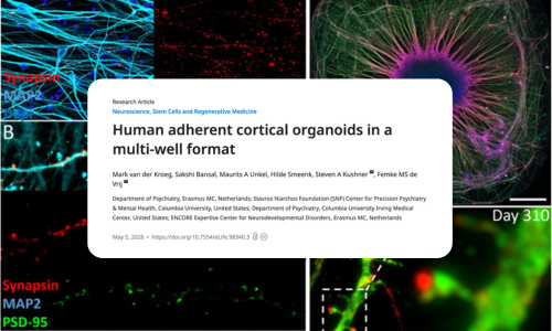
Rather than growing large floating organoids, our team at Erasmus MC Rotterdam developed adherent cortical organoids (ACOs): thin, self-organizing neural tissues grown directly in standard 384-well plates provided by our partner company 3Brain. The resulting structures are smaller, more reproducible, and capable of remaining active for months — features that make them especially compatible with the broader goals of the 3D-brAIn project.
Rethinking how cortical organoids are grown
For this protocol, Femke de Vrij and Steven Kushner’s team began with human induced pluripotent stem cells (iPSCs) — adult cells reprogrammed into a stem-cell-like state and guided toward becoming neural progenitor cells, the precursors of brain tissue.
Instead of allowing the cells to form irregular floating spheres, they seeded carefully optimized numbers of cells into tiny wells. Over roughly eight weeks, the cells self-organized into cortical-like structures only a few millimeters wide and approximately 0.2 mm thick.
That geometry turned out to matter.
Because the organoids remained relatively thin, they largely avoided the oxygen deprivation that commonly damages larger organoids from the inside out — one of the field’s most persistent technical challenges.
Just as importantly, they developed reproducibly. Around 80% of wells generated a single organized structure, an important improvement in a field where experimental variability has often limited scalability and comparison across datasets.
For 3D-brAIn, this reproducibility is foundational. Building meaningful bio-digital brain models requires neural systems that can be monitored consistently over time and compared systematically across experiments, patients, and analytical pipelines.
Rethinking how cortical organoids are grown
The organoids were not simply remaining alive — they were organizing and maturing.
The tissues developed layered arrangements resembling aspects of early human cortical development and contained multiple neural cell types, including excitatory neurons, inhibitory interneurons, astrocytes, and oligodendrocytes.
Over time, the networks became increasingly complex. The researchers observed synapse formation, branching neuronal structures, and synchronized waves of neuronal activity spreading across the tissue.
In other words, the cells were not only present — they were functioning together as networks.
Perhaps most strikingly, the organoids remained viable for up to 10 months without requiring slicing or specialized interventions to prevent tissue death. Long-term stability like this is especially important for studying neurological and psychiatric disorders that emerge gradually over time rather than over days or weeks.
Building stable and active neural networks
For this protocol, Femke de Vrij and Steven Kushner’s team began with human induced pluripotent stem cells (iPSCs) — adult cells reprogrammed into a stem-cell-like state and guided toward becoming neural progenitor cells, the precursors of brain tissue.
Instead of allowing the cells to form irregular floating spheres, they seeded carefully optimized numbers of cells into tiny wells. Over roughly eight weeks, the cells self-organized into cortical-like structures only a few millimeters wide and approximately 0.2 mm thick.
That geometry turned out to matter.
Because the organoids remained relatively thin, they largely avoided the oxygen deprivation that commonly damages larger organoids from the inside out — one of the field’s most persistent technical challenges.
Just as importantly, they developed reproducibly. Around 80% of wells generated a single organized structure, an important improvement in a field where experimental variability has often limited scalability and comparison across datasets.
For 3D-brAIn, this reproducibility is foundational. Building meaningful bio-digital brain models requires neural systems that can be monitored consistently over time and compared systematically across experiments, patients, and analytical pipelines.
What this means for the future of brain modelling
Across neuroscience, there is growing recognition that the future of human brain modelling will depend not only on biological complexity, but also on standardisation, scalability, and functional readout.
Because the organoids are grown in standardized multiwell plates, they are inherently compatible with automated imaging, longitudinal monitoring, and high-throughput screening workflows. Their geometry also makes them well suited for integration with advanced electrode array technologies designed to capture neural activity across three-dimensional tissues over extended periods.
This adherent organoid format helped lay the groundwork for the broader technological ecosystem that 3D-brAIn is now developing. Building on it, researchers at Erasmus MC Rotterdam and LMU Munich continue to refine human stem-cell-derived brain models that can be integrated with high-resolution 3D microelectrode arrays from 3Brain, capable of recording neural activity across complex tissues over extended periods of time. These large-scale electrophysiological datasets are then analysed using AI-based methods developed by the University of Genoa, helping researchers better understand how human brain networks develop, function, and change in disease.
This opens possibilities not only to study disease mechanisms more systematically, but also to move toward something much more ambitious: patient-specific functional brain models that can be analysed computationally and eventually used to support personalised therapeutic strategies.
But the authors are careful not to overstate the model’s complexity. These organoids do not recreate every feature of the human brain, including the folded cortical architecture seen in vivo, and some cell populations still show variability.
But that restraint is part of what makes the work compelling.
The field is increasingly moving away from the idea that larger and more complex automatically means better. In many contexts, a simpler and more reproducible model can ultimately become far more useful — especially when the goal is to generate robust functional data at scale.
And that may be one of the most important shifts emerging from projects like 3D-brAIn: not simply building miniature brains, but building human brain models that are stable, measurable, and precise enough to become truly informative experimental systems.
Read the full article → https://doi.org/10.7554/eLife.98340.3

